2D ECHOCARDIOGRAM:
What is Echocardiogram?
It is a test that uses ultrasound to examine the heart. It allows for accurate assessment of the heart chambers, walls, valves and the great vessels that enter and exit from the heart.
What is a Doppler study?
It’s special part of the exam to assess blood flow direction and velocity.
What information can we get from echo and Doppler studies?
- Size of the heart chambers • Pumping function of the heart
- Valve function
- Fluid in the pericardium
- Congenital abnormalities
Safety of Echocardiography:
It’s extremely safe. How long does it take? Uncomplicated cases can be done within 20-30 minutes.
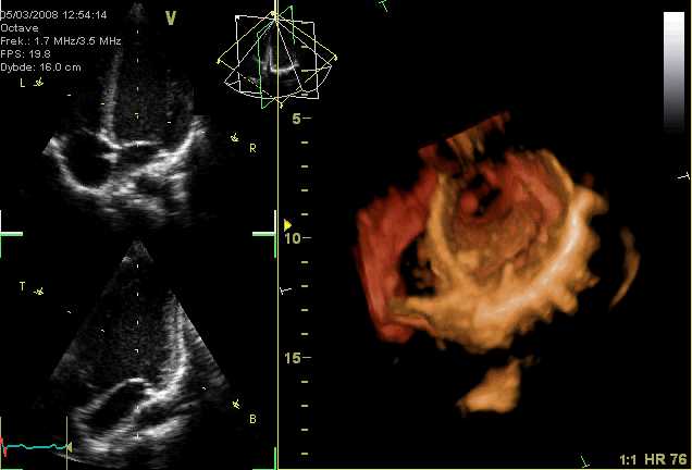
3D ECHOCARDIOGRAM:
3D echocardiography is now possible, using an ultrasound probe with an array of transducers and an appropriate processing system. This enables detailed anatomical assessment of cardiac pathology, particularly valvular defects, and cardiomyopathies.
The ability to slice the virtual heart in infinite planes in an anatomically appropriate manner and to reconstruct Three-dimensional images of anatomic structures make 3D echocardiography unique for the understanding of the congenitally malformed heart.
Real Time 3-Dimensional echocardiography can be used to guide the location of bioptomes during right ventricular endomyocardial biopsies.
Transesophageal Echocardiogram:
This is an alternative way to perform an echocardiogram.
A specialized probe containing an ultrasound transducer at its tip is passed into the patient's esophagus. This allows image and Doppler evaluation which can be recorded. This is known as a transesophageal echocardiogram, or TEE.
Transesophageal echocardiograms are most often utilized when transthoracic images are suboptimal and when a more clear and precise image is needed for assessment.
This test is performed in the presence of a cardiologist, registered nurse, and ultrasound technician.


